Clinical Examination of Knee

Evaluation of Knee:

Evaluation of the Knee Joint includes the following:

  • Complete and Detailed History.
  • Clinical Examination with Special Tests.
  • Correct Imaging.

Complete & Detailed History:

This should include the following:

  • Duration of symptoms e.g pain, swelling, catching / locking
  • Intensity of pain
  • Exact location of pain
  • Type of pain: dull, throbbing, intermittent or continuous
  • Aggravating and relieving factors
  • Popping or clicking noise
  • Inactivity of the muscle
  • Details about previous injury
  • Details about previous surgery

Clinical Examination includes:

Inspection of the Knee

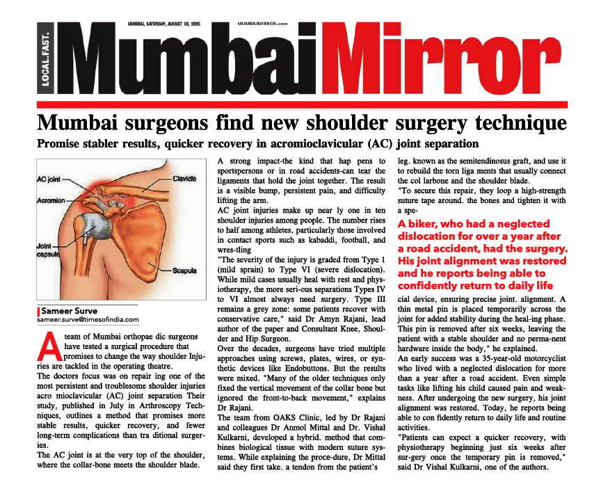
Inspection of the Knee:

Muscle wasting, presence of scars, swelling, redness, & deformity

Palpation of the Knee

Palpation of the Knee includes:

Checking for warmth, location of tenderness, checking for presence of synovitis, checking for presence of effusion.

Measurement of Deformity

Measurement of Deformity:

Muscle wasting, presence of scars,
swelling, redness, & deformity

Range of Motion of Knee Joint

Range of Motion of Knee Joint:

Normal range is from 0 degrees extension to 140 degrees flexion
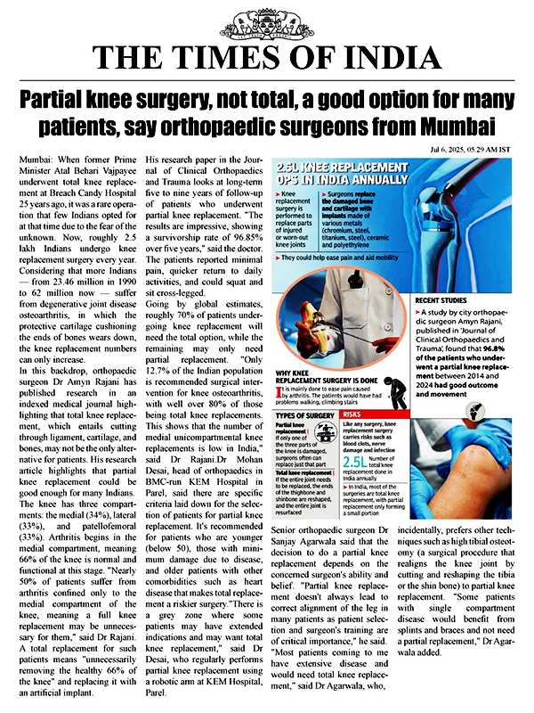
Special Tests:

McMurray's Test for Meniscus Tear:

McMurray's Test for Meniscus Tear

Special Tests:

Lachman’s Test for Anterior Cruciate Ligament:

Lachman's Test for Anterior Cruciate Ligament

Anterior Drawer Test for ACL:

Sag Sign for PCL Tear

Posterior Sag Test for Posterior Cruciate Ligament:

Posterior Sag Test for Posterior Cruciate Ligament

Posterior Drawer Test:

Posterior Drawer Test

Knee Imaging:

X-rays of the Knee:

These are done to diagnose problems in the bones and or joints
The common types of Knee X-rays a Knee Surgeon would require are

Knee (AP Weight-Bearing View)

Knee (AP Weight-Bearing View)

Lateral Knee X-rays

 Lateral Knee X-rays

Axial View of Knee

Axial View

MRI of the Knee:

This is done to diagnose problems with the cartilage, menisci, ligaments or other soft tissues around the Knee.

MRI of Knee

MRI of Knee