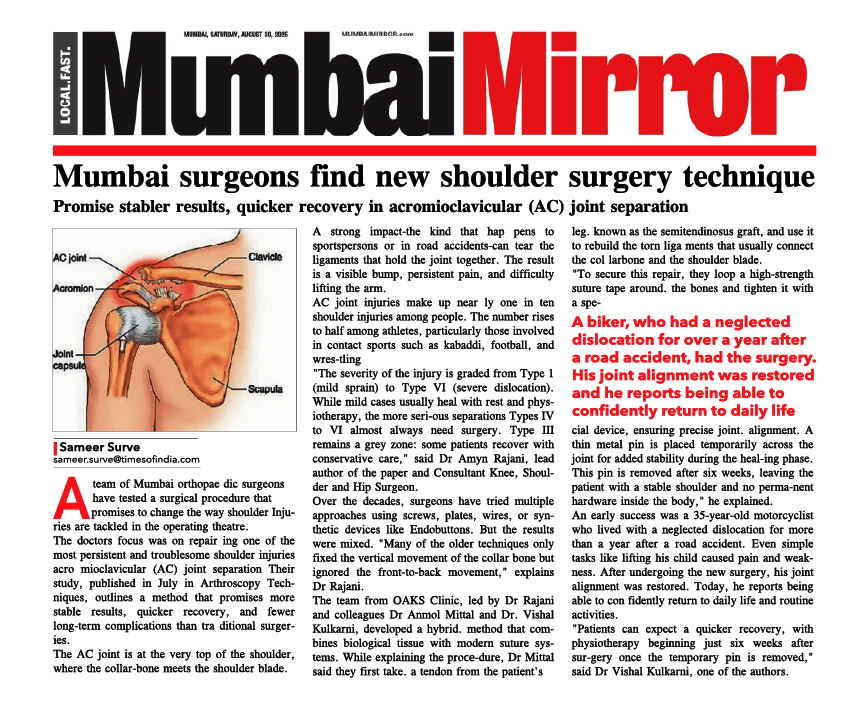
Close Wedge Osteotomy
This surgery was made popular in the year 1950 and is still widely performed in some centers around the world. In a Close Wedge Osteotomy, the surgeon cuts though the tibia on the lateral (outer) side, removes a wedge of bone, and then closes the cut edges together by fixing it with a plate or a staple.
The Close Wedge Osteotomy Surgery involves, making an incision on the lateral side of the Knee to allow the Orthopaedic Surgery Specialists to see the upper end of the tibia. Care is taken to protect the nerves and blood vessels that travel across the Knee Joint.
Once the tibia bone is exposed, two cuts are made through the upper tibia in the shape of a wedge. With the help of x-ray control or image intensifier, correct size of wedge is removed.
The surgeon takes out the wedge, and the two sides of the tibia are brought closer together and held in position with a metal plate, pins or staple. This changes the angle of the tibia and helps straighten the alignment of the Knee. After fixing the two edges of bone with a plate or pins, the surgeon stitches the skin together, and the leg is placed in a padded splint to protect the Knee Joint.
Advantages:
- Better initial stability due to compressive forces
- Immediate weight-bearing
- Quicker union at the Osteotomy site
Disadvantages:
- Shortening of the leg
- Lateral step is created in tibia making future Total Knee Replacements difficult
- Increases the height of patella making patella unstable and increases its chances of subluxation