ACL Tear & Reconstruction
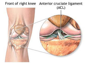
Anterior Cruciate Ligament (ACL):
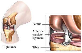
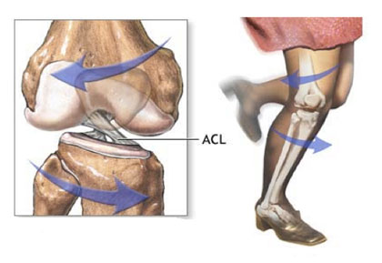
ACL lies within the Knee Joint and is about 4 cm long, binding tibia to femur. The ACL prevents forward slipping of tibia on femur and stops hyperextension of the Knee. It runs through a special notch in femur called Intercondylar Notch and attaches to a special area of tibia called the tibial spine. The ACL is the main controller of tibia’s movement under femur. This is called anterior translation of the tibia. If tibia moves too far, the ACL can rupture.
The ACL is also the first ligament that becomes tight when the Knee is straightened. If the Knee is forced past this point, or hyperextended, the ACL can be torn.
Other parts of the Knee may be injured when the Knee is twisted violently, as in a clipping injury in football. It is also not uncommon to see a tear of Medial Collateral Ligament (MCL) on the inside edge of the Knee and Lateral Meniscus.
Tear of the Anterior Cruciate Ligament:
Mechanism of Tear:
Ruptures involves a sudden deceleration (slowing down or stop), along with a hyperextension, or pivoting in place. Sports-related injuries are the most common.
Structures found in the Posterolateral Knee include Tibia, Fibula, Lateral Femur, Iliotibial Band (IT band), the long and short heads of the Bicepsfemoris Tendon, Fibular (lateral) Collateral Ligament (FCL), Popliteus Tendon, Popliteofibular ligament, Lateral Gastrocnemius Tendon, and Fabellofibular Ligament.
An ACL injury usually and might require ACL Reconstruction Surgery occurs when the Knee is forcefully twisted or hyper extended while the foot remains in contact with the ground.
Numerous types of sports have been associated with ACL tears. Sports which require the foot to be planted and the body to change direction rapidly (such as basketball) carry a high incidence of injury. Football is another sport which gives rise to ACL injury. Football combines the activity of planting the foot and rapidly changing direction and the threat of bodily contact. 80% ACL injuries happen due to non-contact but the remaining 20% happen due to contact related injury for example, a blow to the outside of the Knee when the foot is planted is the most likely contact-related injury.
Symptoms:
Swelling: Your Knee will feel very tight, and will look visibly larger than your uninjured Knee. When both the Knees are put side by side, you will be able to tell if the injured Knee is swollen. Many people describe it as "looking like a grapefruit " - large and puffy. This type of swelling is called an effusion, as it is contained within the joint.
Pain: Pain is very common at initial stage. It is usually described as sharp initially, and then may become throbbing or aching when your Knee swells up. Trying to straighten or bending of the Knee often increases pain.
Feeling or hearing a pop: The pop could be either the ACL Tear or femur and tibia rubbing against each other. Most of the patients will be able to confirm hearing the pop, but some patients may not hear due to anxiety.
Giving way feeling: The Anterior Cruciate Ligament is a major stabilizer of the Knee. It helps the joint communicate with the muscles to keep the Knee stable. If it is disrupted, one may have episodes where the Knee feels like it gives out, or buckles. This is especially true when one walks on it, or tries to turn on the injured leg. This is one of the most common symptoms of a torn ACL.
Sign:
Lachman's Test:
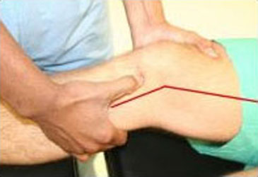
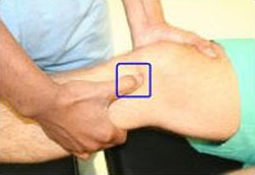
Knee at 30 degree with thumb on Anterior Surface of tibia and give an Anterior Thrust
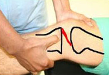
Normal ACL: No Subluxation
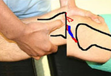
Torn ACL and Subluxation
Anterior Drawer Test:
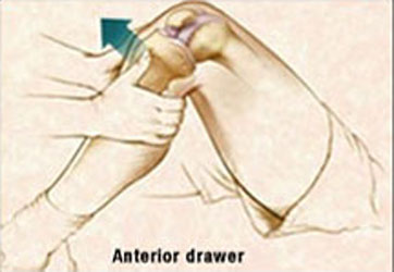

Knee at 90 degrees, sit on the foot of the patient holding tibia with thumbs on anterior surface and pull anteriorly. If there is a tear u will feel the subluxation.
Loss of Range of Motion:
Another one of the symptoms of a torn ACL is decreased range of motion. Most commonly you will have trouble bending it, often unable to bend to 90 degrees. It may also be difficult to completely straighten it out.
Loss of Strength:
Symptoms of a torn ACL also include quadriceps weakness. Due to swelling and injury to the Knee, the quadriceps muscles will be inhibited by the body in order to try to protect the injury. This means that you will have trouble lifting your leg, or straightening it out, both because of pain, and sense a feeling of weakness.
Investigation:
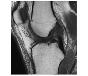
NORMAL ACL MRI
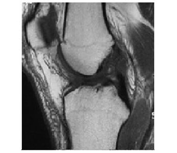
TORN ACL MRI
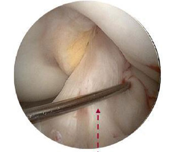
NORMAL ACL
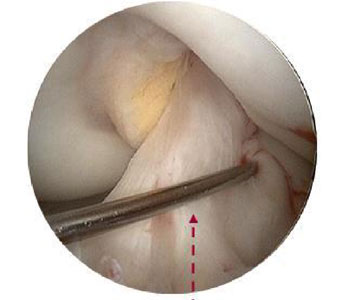
TORN ACL
Immediate steps in treatment of ACL injury:
Irrespective of the fact that surgery is being contemplated or not, the first steps in treatment of an ACL injury are to decrease the swelling via rest, ice compression, elevation (RICE) and anti-inflammatory medicines (ibuprofen, diclofenac, etc.)
Regain full range of motion of the Knee Joint (ability to bend and straighten the Knee all the way). Strengthening the muscle groups (hamstrings and quadriceps muscles) around the knee.
If there are no episodes of buckling or instability, then you may choose not to have surgery. The muscles around the joint can help in giving the knee some stability for activities of daily living. However, this is not usually recommended.
If you plan on being active and participate in sports or if you have ongoing instability episodes then having the ACL reconstructed is an absolute necessity. The ACL reconstruction is an outpatient arthroscopic procedure that involves removing the torn ligament and replacing it with a new ligament (called a “graft”), from you own tissue. (semitendinosus, gracilis).
Arthroscopic ACL Reconstruction:
Do I Need Surgery?
Treatment decisions for ACL tears are always individualized - tailored to each individual. The decision whether to offer ACL Reconstruction Surgery is based on the person's age, activity level, how unstable the Knee is, and whether other structures in the Knee have been injured.
It is important to keep in mind that surgery to reconstruct a torn ACL is not an emergency for most people. Many people with a torn ACL do not need surgery at all. Even though the chances for complete success from surgery are now excellent, surgery is not for everyone. This is because not everyone needs the ligament repaired to return to his or her pre-injury level of function. It is important to distinguish whether the work, recreational, and athletic activities of the person is light, moderate, or strenuous. Another important issue that needs to be understood by the individual considering ACL Reconstruction Surgery is that it requires many weeks and months of hard work in rehabilitation following the reconstruction. This needs commitment and time.
Surgery:
The most common autograft options are bone-patellar-bone tendon and hamstrings. ACL Reconstruction Surgery is typically done as an outpatient procedure. Depending on graft choice, open incisions may be necessary to harvest the tissue that is to be used as the new ACL. Knee Arthroscopy is then performed to inspect the Knee, treat additional injuries (meniscus tears or cartilage damage), and to prepare the Knee for the new ACL.
Once the graft tissue has been prepared and the torn ACL tissue has been removed, the surgeon is ready to place the ligament within the Knee. Small tunnels (7-10 mm) are drilled in the tibia and femur to allow the ligament to be pulled up into the Knee.
Accurate placement of these tunnels is critical to successful ACL Reconstruction Surgery. After the ACL graft is in position, fixation devices (screws, washers, buttons, etc.) get used to keep it there until it can heal into its place.
Surgical steps:
- The orthopaedic surgeon inspects the Knee and removes the remains of the old ACL using an arthroscopic shaver.
- The graft which gets used for reconstruction, is harvested and prepared for the replacement. Usually the patellar tendon or the Semitendinosis and Gracilis tendon autografts are used in athletes.
- After harvesting the tissue, a hole is drilled from the front of tibia diagonally into the Knee and ends up where the ACL attaches to the top of the shin. Next, the orthopaedic surgeon drills a hole in femur at the anatomical foot-print on the lateral femoral condyle.
- The harvested replacement graft pulled into place through the holes which were just drilled and locked after flipping the Endo-CL Button.
- The new ligament is then held into place by two bio-absorbable screws or metallic screws.
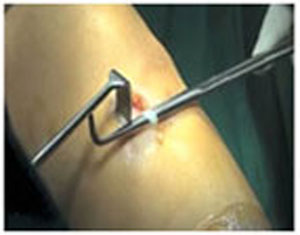
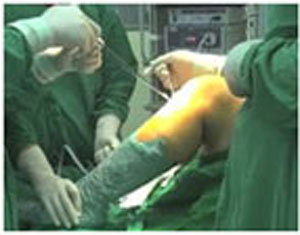
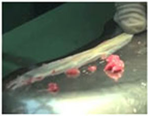
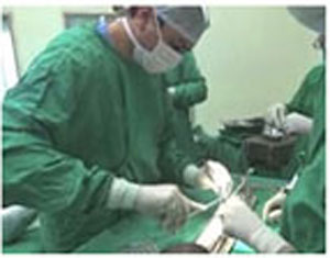
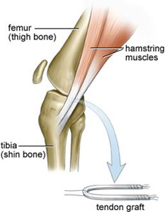
Graft Harvesting
Graft Preparation
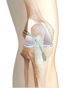
Final Fixation
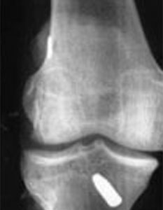
Post-Op X-ray
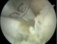
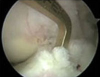
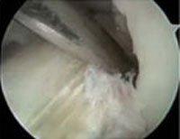
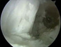
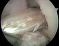
In this procedure, rather than using the patellar tendon, the surgeon uses the patient's own hamstring tendon, either the Semitendinosus and Gracilis tendons from the same leg.
There are several variations of this technique. Newer hamstring fixation techniques have been developed to match and even exceed the initial pullout strength of the patella bone tendon bone procedure. Special screws with threads designed not to cut the hamstring tendons are able to fix the tendon within the bone tunnel, as described with the patella bone tendon bone technique.

