Do you require Knee Arthroscopy?
If you have swelling, persistent pain, catching, giving way, and loss of confidence in your Knee and conservative measures such as regular use of medications, Knee supports, and physical therapy, have provided minimal or no improvement, you may benefit from Arthroscopic Knee Surgery.
Conditions which can be diagnosed and treated by Knee Arthroscopy.
Indications for Arthroscopic Knee Surgery:
- Loose Body
- Synovitis
- Meniscus Tear
- Cruciate Ligament Tear
- Cartilage Lesions
Meniscus:
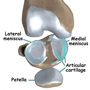
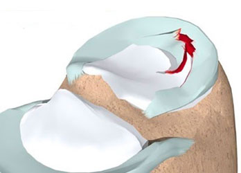
There are two menisci in the Knee (Medial & Lateral), which act like gasket to distribute weight from femur to tibia. They in turn also protect the Articular Cartilage.
What is Meniscus Tear?
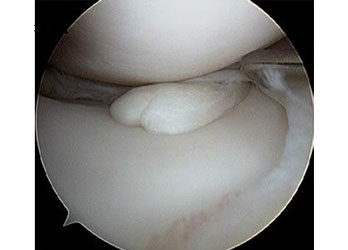
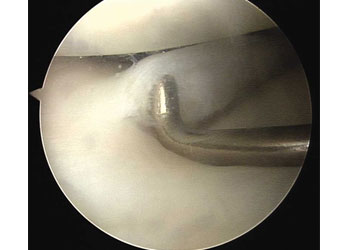
Tear in the meniscus can lead to pain, swelling, locking. Long term effects of torn meniscus due to constant rubbing on Articular Cartilage can lead to Osteoarthritis.
What are the symptoms of a Torn Meniscus in your Knee?
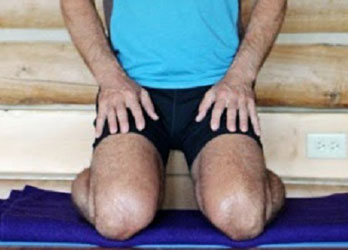
- Extreme pain, especially when the area in the knee is touched
- Swelling
- A popping sensation in the knee
- Difficulty in performing some of the simplest joint functions
- Difficulty in bending and straightening of the knee
- Problem in moving your knee
Causes of Meniscus Tear in Knee
- A wrong turn and squat while sprinting or performing long jump and high jump and you might badly affect and tear the meniscus in your knee.
- People above 60 years of age usually suffer from degenerative tears- the reason: their cartilage weakens and becomes thinner over time.
- Untreated meniscus tears can badly impact your ability to perform daily activities and you may have problem in performing some of the simplest tasks. In some cases, it can develop into serious long-term knee problems like arthritis. Torn meniscus can also pull the fragments of the cartilage of your knee joint.
What are the Meniscus Tear Treatment Options?
Treatment for meniscus tears depend on the size, where is it located within the knee cartilage and most importantly what type of tear it is that your knee is affected by. If the tear is not that major then initially you will be advised to opt for conservative treatments. Icing, resting are some of the treatments that you will be asked to try. If these conservative treatments don't work then arthroscopic knee surgery is the best option for your torn and wormed out meniscus.
What is a Meniscus Repair?
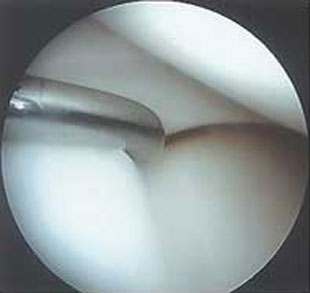
Your Arthroscopic knee surgeon will make small cuts in the knee. He will insert an arthroscope which will allow him to have a good look at the meniscus tear and operate inside the knee joint.
What is Meniscectomy?
A Meniscectomy surgery is performed to remove torn part of your meniscus.
What is a Meniscus Root Repair?
A meniscus root repair involves reattaching the meniscus back to bone where it has been torn. The steps in a meniscus root repair are very intricate and our biomechanical studies have validated that the steps are essential.
What is a Cartilage Repair?
An Arthroscopic Knee Surgery which involves the restoration of the damaged hyaline cartilage in your knee joint.
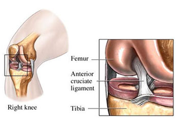
Cruciate ligaments:
Anterior Cruciate Ligament: Provides Anterior Stability to Knee Joint.
Posterior Cruciate Ligament: Provides Posterior Stability to Knee Joint.
Tear of Cruciate ligaments:
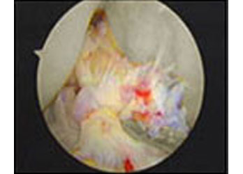
Tear in ACL and or PCL can lead to pain, swelling, and instability of Knee Joint. Due to the tear a feeling of give way occurs whilst changing direction.
Diagnosis confirmation: MRI
Treatment:
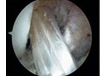
A tendinous graft is harvested from semitendinosus muscle and or gracilis muscle and is replaced in place of original cruciate
Patellofemoral Joint Issues:
- The common Patellofemoral issues are problems due to maltracking, malalignment, Chondromalacia Patella & osteoarthritis of Patellofemoral Joint.
- Symptoms are those of feeling of patella slipping away, pain and swelling.
- Chondromalacia, maltracking due to soft tissue imbalance and many other Patellofemoral issues can be dealt with by Knee Arthroscopy.
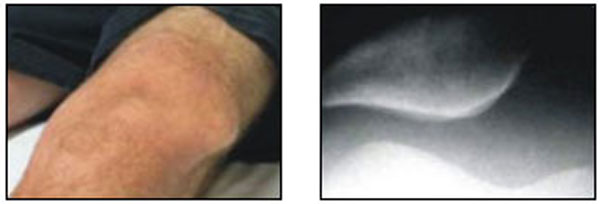
Before Surgery
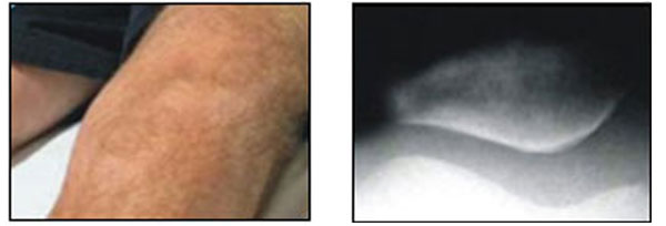
After Surgery
Synovium:
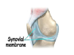
The normal Knee Joint is surrounded by a membrane (the Synovium) which produces a small amount of thick fluid (Synovial Fluid). The synovial fluid helps to nourish the cartilage and keeps it slippery. The Synovium also has a tough outer layer (the joint capsule) which protects & supports the joint.
Synovitis:
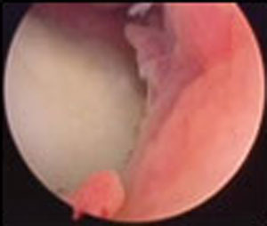
Inflammation of the Synovium is called Synovitis. This may occur due to rheumatoid arthritis, infections, trauma or injury, autoimmune. Symptoms are swelling, pain and stiffness.
Treatment:
Synovial Biopsy and medical management or subtotal Synovectomy.
Localised Articular Cartilage Lesions in the Knee Joint:
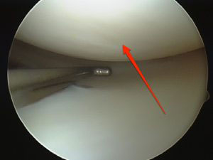
Normal Articular Cartilage:
The ends of the long bones are covered with a smooth white glistening surface called the Articular Cartilage. The Articular Cartilage helps in joint motion by lubrication and by preventing friction.
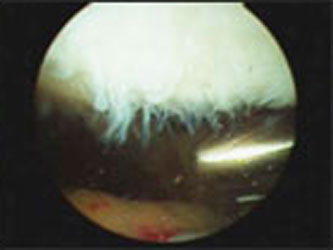
Traumatic events can damage Cartilage, and as we age, the biology of the Cartilage changes, predisposing it to wear and injury.
Treatment of Articular Cartilage Defects:
Arthroscopic Chondroplasty:
Loose cartilage flaps and debris within the Knee are removed using an Arthroscopic shaver or a radiofrequency ablator.
Mosaicplasty:
Small focal lesions can be treated with Mosaicplasty In which cylinder pegs of Osteochondral Grafts are taken from non-weight-bearing portions of the Knee and transferred to the cartilage defects in the weight -bearing portion.
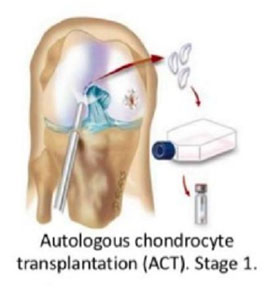
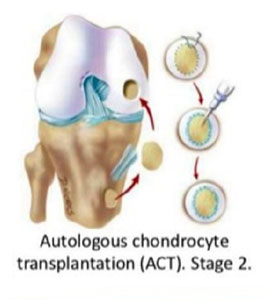
Autologous Chondrocyte Implantation: (ACI)
This is a two-stage procedure. The first stage involves Arthroscopic Biopsy of normal cartilage from non-weight-bearing area of Knee Joint. The biopsy is then sent to cartilage expansion laboratory where the cells are cultured and multiplied. It takes about 6 weeks for the multiplication of cells. The second stage involves the implantation of these cartilage cells in the damaged area. This is done by an open procedure.
