What is High Tibial Osteotomy (HTO)?

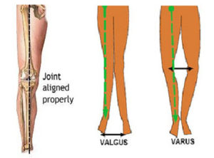
In a normal Knee the mechanical axis of the limb, should pass through the center of the Knee.
In a Varus Knee, the mechanical axis of the limb passes medially (inner side) to the center of the Knee.
In a Valgus Knee the mechanical axis passes laterally (outer side) to the center of the Knee.
The Knee is divided into three halves, or compartments. The medial compartment is the inside half of the Knee and is formed by the articulation of the medial femoral condyle and the medial tibial plateau. The lateral compartment is the outside half of the Knee and is formed by the articulation of the lateral femoral condyle and the lateral tibial plateau. The third compartment is the patellofemoral compartment which is formed by articulation between the femoral trochlea and the patella.
Osteotomy (osteo-bone, tomy-cutting) means cutting of the bone. This is one of the oldest orthopaedic procedures with excellent functional outcomes. Osteotomy is either done to correct a deformity in the bone and/or to correct the mechanical axis of the lower limb in cases of arthritis of the knee joint in patients with bow legs or Knock Knees. In bow legs or Knock Knees with arthritis, the osteotomy is usually done at the level of upper tibia and hence the name High Tibial Osteotomy Surgery.
The surgical aim of High Tibial Osteotomy is to correct the mechanical axis of the lower limb thus redistributing the weight away from the damaged, towards the undamaged compartment. The damaged compartment is consequently offloaded, which in principle reduces the progression of surface wear and buys time before joint replacement surgery might be required. One has to be careful however not to overcorrect the alignment too much, as this might lead to increased pressure and subsequent osteoarthritis in the otherwise normal compartment.
As there have been excellent results with Total Knee Replacement Surgery, the use of High Tibial Osteotomy by surgeons is diminishing. In young people the Knee usually wears out either on the inner side or the outside producing a bow leg or Knock Knee Deformity. Realigning the angle made between the bones of the leg can shift your body weight so that the healthy side of the Knee Joint takes more of the stress. This will give symptomatic relief from pain as well as prevent further joint damage.
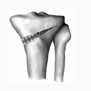
This procedure is now ideally indicated for young patients with osteoarthritis only in one compartment of the tibiofemoral joint. There are two methods to realign the Knee Joint. One involves taking out a wedge of bone; the other involves adding a wedge of bone. In a closing wedge osteotomy, the surgeon cuts through the tibia on the lateral (outer) side, removes a wedge of bone, and pins the open edges together. In an Opening Wedge Osteotomy, the surgeon cuts through the tibia on the medial (inner) side and opens a wedge, adding a bit of bone graft to hold the wedge open. High Tibial Osteotomy can be used in conjunction with arthroscopic cartilage procedures to give better results.

