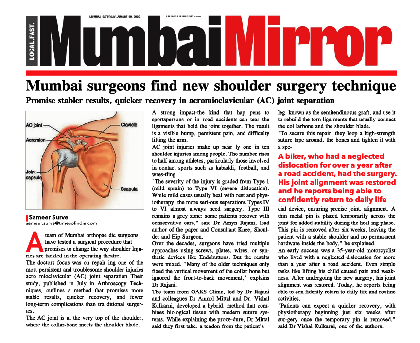
Articular Damage & Arthroscopic Articular Cartilage Repair

The ends of the long bones are covered with a smooth white glistening surface called the articular cartilage. The Articular Cartilage helps in joint motion by lubrication and by preventing friction. This slick surface is designed to minimize pressure and friction as you move.
Cartilage tissues are devoid of any nerves. They also lack supply of blood or lymph vessels, which normally nourish other parts of the body. Without a direct supply of nourishment, cartilage is not able to heal itself, if it gets injured.
Injury or lesions of the cartilage can be superficial or deep. These rough spots or holes in the cartilage can damage the design of the joint, due to which the Knee Joint Pain becomes inflamed and painful. If the injury, or lesion, is large enough, the bone below the cartilage loses protection, and pressure and strain on this unprotected portion of the bone can also become a source of Knee Joint Pain. Loose pieces of the cartilage act like foreign body and can cause locking and catching of the joint.

Grades Of Articular Cartilage Lesions
Cartilage lesion of the knee are classified from I (one) to IV (four) depending upon the depth of the cartilage involved and the severity of the lesion. In grade I lesions, the cartilage has a soft spot. Grade II lesions show minor tears in the surface of the cartilage. Grade III lesions have deep crevices. In grade IV lesions, the tear goes all the way up to the underlying bone.
The following images show each type of defect:


The grade IV lesion goes completely through all layers of the cartilage. It is diagnosed as a full-thickness lesion. Sometimes part of the torn cartilage breaks off inside the joint and begins to move around within the joint, causing even more damage to the surface of the cartilage.
Symptoms:
swelling, pain, catching or locking, stiffness, decreased range of motion
Diagnosis:


Management:
Treatment of Articular Cartilage lesions is dependent on multiple factors. Patient age, activity level, and mechanism of injury commonly dictate management. Young patients with traumatic injuries are treated aggressively to try to restore a normal articular surface within the Knee.
Conservative Knee Arthroscopic Treatment is indicated for patients who are present with diffuse osteoarthritis. Such patients are treated by physical therapy, medications activity modification, orthotics, bracing, and injections. Failure of conservative management may be an indication for surgical intervention, depending on the patient’s symptomatology.
The size of the lesion, the quality of the bone behind the lesion, and the Arthroscopic Knee Surgeon preference are some of the factors considered before choosing the appropriate surgical procedure for a particular lesion.
Surgical Procedures For Cartilage Lesions:
Arthroscopic Abrasion Chondroplasty:
This Arthroscopic Knee Surgery procedure includes removal of loose chondral flaps and smoothing of the Articular Cartilage (chondroplasty). It is utilized for small areas of superficial cartilage damage in patients that experience joint pain and have associated mechanical symptoms. The goal of Arthroscopic Articular Cartilage Surgery is to eliminate catching and locking from these loose pieces of cartilage and to try to prevent propagation of these chondral flaps. The scraping action causes a healing response in the bone. In time new blood vessels enter the area and fill it with scar tissue (Fibrocartilage) that is like Articular Cartilage.


Arthroscopic Microfracture:
< 2 cm) of cartilage damage that are surrounded by healthy tissue. There is presence of rich blood supply in the bone beneath the cartilage. Microfracture procedures try to access this osseous vascular supply in order to heal cartilage defects. In this procedure small holes are created in the bone at the base of a cartilage lesion. This allows cells from the bone marrow to fill in the defect and stimulate new cartilage growth. This new cartilage is a scar-type of cartilage called fibrocartilage. Wide spread cartilage treatments are a contraindication to this type of management.
Arthroscopic Mosaicplasty:
In this procedure the Cartilage defects are appropriately sized plugs osteochondral grafts are obtained from the non-weight-bearing portion (usually on the top and outside border) of the Knee cartilage.
The harvested plugs are then transferred to the cartilage defects in the weight-bearing portion of the Knee. This procedure is done for lesions which are < 2cm, hence cannot be done in patients with diffuse cartilage damage.


Lesion

Donar-Site

Pegs

After-Implantation