When it comes to diagnosing synovial lesions in the knee, orthopedic surgeons often rely on MRI scans and arthroscopy to guide clinical decisions. While these tools are advanced and widely used, a critical study published in the Journal of Experimental Orthopaedics reveals that relying solely on imaging can lead to misdiagnosis—and mistreatment.
In this blog post, we explore three revealing case studies that underscore the importance of histopathological examination in accurately diagnosing synovial knee conditions.
Understanding Synovial Lesions of the Knee
The synovium is the soft tissue lining of the joints. Inflammatory or abnormal growths in this lining can cause:
- Knee pain
- Swelling
- Limited range of motion
Commonly suspected conditions include:
- Lipoma arborescens (fatty villous synovial growth)
- Synovial chondromatosis
- Pigmented villonodular synovitis (PVNS)
- Chronic inflammatory synovitis
These conditions often appear similar on MRI and during arthroscopy, making clinical diagnosis challenging.
Case Studies: What the Research Revealed
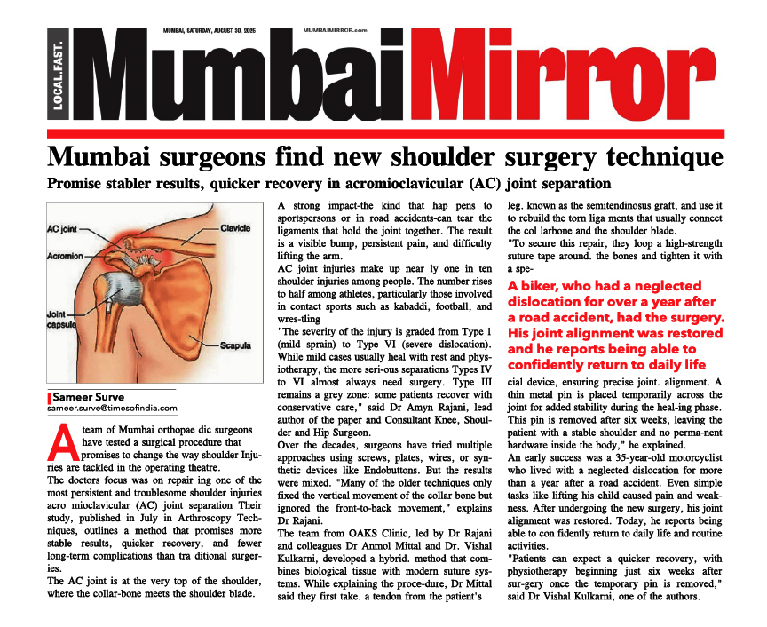
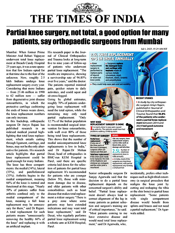
A team led by Dr. Amyn Rajani a leading Consultant Knee, Shoulder & Hip Surgeon in Mumbai and colleagues reviewed three cases where knee swelling and MRI suggested classic synovial lesions—but histopathology told a different story.
🩺 Case 1 & Case 2:
- MRI Diagnosis: Lipoma arborescens
- Arthroscopy Findings: Confirmed villous fat-like projections
- Final Diagnosis via Histopathology: Chronic inflammatory synovitis
- Treatment: Rheumatologic care with methotrexate and hydroxychloroquine
- Outcome: Complete resolution of symptoms without further surgery
🩺 Case 3:
- MRI Suspicion: Loose chondral body
- Arthroscopy: Confirmed mobile intra-articular mass
- Histopathology: Organized hematoma, not cartilage
- Outcome: No recurrence after removal
Key Takeaways for Orthopaedic Surgeons
|
Insight
|
Why It Matters
|
|
MRI & Arthroscopy are not always conclusive
|
They can mislead diagnosis, especially in inflammatory conditions
|
|
Histopathology is crucial
|
Only microscopic analysis can confirm the exact nature of synovial lesions
|
|
Treatment plans can drastically change
|
From unnecessary surgery to effective medical therapy
|
|
Early diagnosis improves outcomes
|
Prevents joint damage and chronic inflammation
|
The Role of Histopathology in Knee Diagnosis
This study strongly advocates incorporating routine synovial biopsy and histopathological examination into knee arthroscopy, especially when:
- Lesions are atypical or persistent
- MRI findings are inconclusive
- There's a risk of mistaking inflammatory conditions for structural issues
Improving Diagnosis Through Multidisciplinary Care
In each of the cases, collaboration with rheumatology specialists was crucial to treatment success. This highlights the importance of:
- Cross-specialty referrals
- Personalized care planning
- Monitoring autoimmune or systemic causes of knee inflammation
Conclusion
This paper reminds us that “It’s not always what it looks like” when diagnosing synovial lesions of the knee. Even advanced imaging techniques like MRI and arthroscopy may fail to differentiate between benign growths and inflammatory conditions.
Surgeons should consider histopathological confirmation as a gold standard—especially when clinical and imaging findings don’t fully align.
If you're experiencing unexplained knee pain and swelling, or if you're a clinician managing complex knee cases, remember: accurate diagnosis leads to better outcomes.